Melissa Valdellon

Patient case for July 21, 2023

Today’s case is a short one. The patient is a 38 year old Hispanic female who was last seen a year and a half ago for a diabetes eye exam. She had been correctable to 20/20-3 and 20/20-1 with a refraction of OD +0.25-2.00×005, OS plano DS. She comes in today saying she has blurred distance vision especially when driving at night and that she never filled the glasses prescription from last year and has never worn glasses ever.

As I mentioned, she has had diabetes since about 2003 with a latest HbA1c of 10.1 from 3 months ago. She also has diagnoses of anxiety and depression, migraine, vertigo, gastritis, and reports quitting smoking about 3 weeks prior. Her medications include albuterol, zyrtec and flonase, glipizide, metformin, and protonix. She has an allergy to eletriptan (she gets hives).

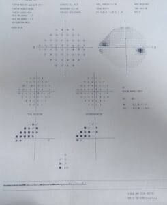
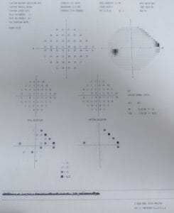
Confrontation testing of pupils, EOMs, visual fields are all normal. Incoming uncorrected visual acuity is OD 20/50-2, OS 20/25-2.

Retinoscopy shows OD plano -2.00 x 180, OS plano -0.50 x 180

Refraction shows OD -0.75 -1.25 x 175  20/25, OS -0.50 DS 20/25+2

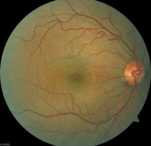
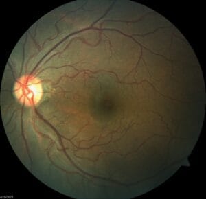
Anterior segment shows some mild punctate erosions inferiorly of both eyes and is otherwise clear with IOPs of 13/13 mm Hg. Posterior segment of both eyes is clear of any pathology or DM retinopathy.

What would you prescribe for this patient? Do her visual acuities match her refraction results?

She had a myopic shift in her eyes compared to her last exam 1.5 years ago. Are you more or less willing to prescribe her glasses because of this shift?

The patient also reports her history of vertigo. Does this mean you should modify her prescription in any way? Considering she’s also never worn glasses in the past, would you modify her prescription any differently?

Let me know your thoughts!

Patient case for July 14, 2023

And now for something a little different. Today, let’s talk about a 47 year old Hispanic male who came in for a second visit. We first saw the patient 2 months prior for his diabetes eye exam. At that visit, he had reported blurred distance vision, OD>OS, and he doesn’t normally wear glasses.

He’s had DM2 for about 11 years and his latest HbA1c was 6.3 in March of this year. Systemically, he also has hyperlipidemia. He takes Flonase and Claritin as needed for seasonal allergies, metformin for his diabetes, and simvastatin for the hyperlipidemia. He has no allergies to medications.

Pupils, EOMs, confrontation fields are all normal. He is correctable to 20/20 with a mild prescription in each eye. Anterior segment is unremarkable and IOPs are 15/17 mm Hg OD/OS, respectively. Posterior segment findings are in the photos below:

As you can see, there’s no evidence of diabetic retinopathy, but there’s a significant hemorrhage on the rim of the optic disc of the right eye.

Given that this patient is only 47 years old, is this optic nerve hemorrhage likely related to glaucoma or not? What are some other causes of optic nerve hemorrhages? Does he fit the profile for any of these other differentials?

At the second visit, we obtained the following information:

Pachy: 619/620
Gonioscopy: CB 360 with 2+ pigment and normal approach OU
IOP 16/14 mm Hg
HVF 24-2:

Based on all the data you have after these two visits, now do you think this patient has glaucoma or no? How would you manage this patient? I’m looking forward to a lively discussion here. Thanks!

Patient case for July 7, 2023

I know we’ve been doing a lot of pediatric prescription cases lately (sorry, not sorry?), but here’s another one to get you thinking.

The patient is an 11 year old Hispanic female coming in for her first eye exam after failing a vision screening with her pediatrician. The patient reports that vision is blurry both distance and up close. She is otherwise healthy, has no allergies, and is not taking any medications.

Distance VA is OD 20/30-2, OS 20/40, and pinholes to 20/25 in each eye. Also, near VA is 0.4/0.5M-1 in each eye.

At this point, can you anticipate what kind of refractive error you’d expect here? Take a guess and keep those numbers handy.

Pupils, EOMs, confrontation fields are all normal. Cover test is ortho distance and near.

Auto-refraction results show:
OD: +0.75 -0.50 x 139
OS +1.25 -0.25 x 098

Refraction, however, yields you this:
OD: -0.75 -0.50 x 005  20/25
OS: -1.250 DS  20/30

Now what? The child is getting tired and annoyed at being here and she’s not feeling like she’s seeing dramatically better compared to when she first came in. What other testing should you do?

While the patient herself did not mention any headaches or eyestrain, a clue that this patient could have more binocular vision problems is the fact that her near visual acuities were also reduced. A minor amount of myopia or astigmatism really shouldn’t be enough to lose a whole line of acuity at near.

So we ended up checking accommodation. NPA on repetition was 8D, 7D, 5D with both eyes open. And after all that fatigue, NPC was 12cm. What are the patient’s age based norms for NPA and NPC?

We next dilated the patient with tropicamide and phenylephrine. Was cyclopentolate indicated for this exam? Would it have been wrong to use it?

Ocular health was unremarkable for both eyes.

Damp auto-refraction results were:
OD +1.25 -1.25 x 007
OS: +1.50 -2.50 x 171

What would you prescribe for this patient? What conditions are affecting this patient that you would discuss with mom about? And of course, what’s your follow up plan? Chime in below!

Patient case for June 30, 2023

Today, let’s discuss this pediatric patient who came into the office for their first eye exam in the states after immigrating from Turkey. A previous eye doctor had let him know he had an eye turn and was given glasses. Mom says that the glasses helped the eye turn, but then the glasses were lost sometime in the last month. The patient is otherwise healthy, has no allergies, and is not taking any medications.

Pupils, confrontation fields, EOMs, and color vision are all normal. He did not appreciate randot stereoacuity and he reported 5 lights on worth 4 light.

Distance cover test reveals about 27pd left esotropia, whlie at near, it’s measured as 16pd left esotropia; the patient was uncorrected for both emasurements.

Uncorrected visual acuity is OD 20/20, OS 20/20.

Dry retinoscopy and refraction show:
OD: +0.75 DS  20/20-
OS: +1.25 -0.50 x 175  20/20

At this point, what drops would you use to dilate this patient and why? Can you review the different properties and effects of Tropicamide vs Cyclopentolate vs Phenylephrine?

~ ~ ~

We ended up using 1% Cyclopentolate for this patient and got the following results:
OD: +3.00 -0.50 x 180  20/20
OS: +3.75 – 0.50 x 170  20/20

All ocular health findings were normal.

What glasses prescription would you recommend for this patient? Given that the patient is 8 years old and able to see 20/20, what is your recommended follow up for this patient? If the patient were actually 5 years old, would any part of your management be different? And lastly, what are your thoughts around strabismus surgery in this case?

I’m looking forward to reading your thoughts.

Patient case for June 23, 2023

I am very grateful to work in a community clinic where I can speak with fellow doctors of different specialties about our mutual patients and they feel welcome to walk over anytime they want to discuss one of their patients with any eye concerns. This following case is just one recent example.

One of the clinic pediatricians came by asking if I could see one of her patients. The patient is a 13 year old female complaining of a sudden onset red right eye that she noticed when she first noticed upon waking up four days ago. Besides the redness, she says it felt like something was in her eye and irritating it. She reports that the eye has been watery with some discharge. She doesn’t remember anything getting into her eye, but she says she used some shimmery makeup two days prior and thinks maybe that could have irritated it.

After a day of no improvement, she went to the emergency room and was prescribed polytrim antibiotic eye drop. She reports good compliance with using the medication every 3 hours as prescribed but there has been no change in the redness or irritation and now the redness is starting to affect the left eye, though not as bad yet.

Overall, she says her eyes feel itchy and they burn. They don’t hurt really but she does have a small headache. And on further questioning, she reports that she hasn’t been recently sick and no one in her family and no one she’s been around in the last few days has been ill either.

Medically, the patient is very health with no conditions, no allergies, and is not taking any other medications besides polytrim eye drop.

Incoming visual acuity is 20/20-3 and 20/25+2 with easy pinhole to 20/20 each eye. Pupils and EOMs are normal both eyes.

Anterior segment exam findings shows clear lids and lashes, but conjunctival hyperemia, chemosis, mixed papillae and follicles OD worse than OS. There is patchy 1+ PEE peripherally on the cornea OD but otherwise clear centrally and no PEE in the left eye. Anterior chambers are deep and quiet, irides are brown and flat, lenses are clear, and vitreous is normal in both eyes. A small pupil posterior segment evaluation shows pink, distinct nerves with 0.40 cupping both eyes, shiny RNFL, normal vasculature, and flat and even macula of both eyes. IOPs are equal at 12/12 with Goldmann tonometry.

Given what you know, does this match the ER diagnosis of bacterial conjunctivitis? What in the exam findings suggest that this is bacterial in nature and what doesn’t?

What else is on your differential diagnosis list? How can you rule in or out allergic conjunctivitis? Viral? Iritis or uveitis? Do you think the patient’s headache is related to her ocular symptoms or not? What about her eye makeup use two days prior?

I can’t wait to read your thoughts!

Patient case for June 19, 2023

Today’s patient is a 35 year old Hispanic male who is coming in for his annual exam. He is reporting some increased mild blur in his glasses, which he did not bring into the exam. He also wants to try contact lenses. He is otherwise healthy, has no allergies, and is not taking any medications.

Incoming visual acuity is OD 20/60, OS 20/50. Pupils, EOMs, and confrontation fields are all normal in both eyes.

Refraction results are:
OD: plano -4.00 x 017   20/20-
OS: -0.25 -2.75 x 159   20/20

Refraction results from two years ago showed 0.50D less cyl in the right eye but otherwise everything else was stable.

Anterior segment is clear and normal in both eyes, IOPs are 16/16, and posterior segment is normal with small nerves and small cups of 0.20 round OU, clear posterior poles, and stable lattice degeneration in each eye compared to 2 years prior.

How would you counsel this patient in terms of their wanting contact lenses? What would be your first choice to try? Is there additional testing you want to perform before trying lenses? Is there any hesitation recommendation contact lenses given he’ll be 40 in just a few more years?

Patient case for June 13, 2023

A 62 year old Hispanic female came in for her annual DM eye exam and was complaining of blurred vision after losing her glasses in Mexico a few months ago.

Medically, she is taking medications for mixed hyperlipidemia, osteoarthritis, type 2 DM, gastroesophageal reflux disease, and POAG with reported good compliance for everything. Her latest HbA1C was 7.4 and she did not check her blood sugar the day of her exam.

Incoming uncorrected visual acuity was 20/100 and 20/300 with pinhole to 20/30 and 20/50. Pupils and EOMs were normal.

Refraction results showed:
OD -22.5 DS  2025
OS -3.00 -0.50 x 170  20/40

Anterior segment showed meibomian gland dysfunction, nasal and temporal pterygium, 2-3+ NS, and vitreous syneresis, all in both eyes, though the cataract may have been slightly more hazy in the left than the right. IOP today was 14/14.

Posterior segment showed thin superior and inferior rims of both optic nerves with C/Ds of 0.65. There had been a hemorrhage on the inferior rim of the left optic nerve back in 2021. That has since resolved but there is now a corresponding mild nerve fiber layer defect. Otherwise, posterior segment was clear and without evidence of diabetic retinopathy.

For more background, this patient has been monitored for her nerve appearance since 2018. Pachymetry is 538/550. Gonioscopy is open in both eyes with no angle recession.

The patient has done multiple visual fields and for the right eye, there is a possible inferior nasal step that “comes and goes”, showing up in some fields but not all. The left eye also shows an inconsistent superior nasal step defect.

OCTs of the nerves show superior thinning that had been stable since 2018, but progressive inferior temporal thinning in the left eye more than the right since 2018.

Treatment of Latanoprost QHS was initiated in 2021 when the optic nerve hemorrhage was first seen. Prior to treatment, IOPs ranged from 19-22 OU. After treatment, her IOPs have fluctuated from 14-22, achieving IOPs of 15 or lower in half the visits since treatment was initiated.

Sorry for the lack of imaging to go along with the case but hopefully you were able to follow along. What would you prescribe for this patient for their refractive error or how would you counsel them in regards to their vision? What would you tell the patient and their doctor about their diabetes management? And what would you like to do about this patient’s glaucoma?

I look forward to hearing your thoughts.

Patient case for June 5, 2023

A 27 year old Hispanic female came in for her comprehensive eye exam, complaining of blurred distance and near vision without glasses. Her last eye exam was a year ago elsewhere – she was not prescribed anything at that time.

Her medical history includes irritable bowel syndrome, depression, and migraines. She takes amitriptyline, sumatriptan, trazadone, and is using a birth control implant.

Incoming vision is OD 20/20-1, OS 20/25-3. Pupils and EOMs are normal.

Retinoscopy is plano in both eyes with visual acuity OD 20/20, OS 20/40+1. Refraction doesn’t yield any change for the left eye and it seems to be fluctuating throughout refraction.

Anterior segment is clear before drops, IOPs are 17 mmHg, 15 mmHg OD/OS, respectively. Posterior segment is clear with moderate cups of 0.65 round OD, 0.60 round OS and a vitreoretinal tuft in the left eye. Another look at anterior segment shows diffuse epithelium disruption left eye and clear right eye after dilation.

Auto-refractor after dilation shows:
OD: -0.25-0.75×173  20/20-1
OS: -1.25 -0.75×164  20/30

Retinoscopy after dilation shows:
OD: +1.50-0.50×180
OS: +1.00-0.50×035

What are your differentials for the decreased vision in the left eye? What extra tests would you like to perform to rule your differentials in or out? How would you manage and counsel this patient at the end of today’s exam based on your different differentials?

I look forward to hearing your thoughts.

Patient case for May 30, 2023

For today’s case, let’s talk about Bell’s Palsy.

A 50  year old Hispanic female came in with a complaint of vision changing on the left side for the last 1.5 months. She is currently using OTC +2.75 reading glasses, which aren’t working well. She also reports more photophobia, watering, and dull pain on the left side. She was diagnosed with Bell’s Palsy on the left side of her face since mid-February. Health-wise, there is nothing else going on and she is not taking any medications.

Uncorrected visual acuities are OD 20/20, OS 20/40 with no improvement on pinhole, retinoscopy, or refraction for the left eye. Confrontation  testing of pupils, EOMs, and fields are normal. IOPs are OD 17 mmHg, OS 16 mmHg.

Anterior segment findings show mild and equal mild meibomian gland dysfuction, 1+ NS, and vitreous syneresis of both eyes. In addition, the left eye shows a mild papillary response and significant NaFl staining response on the cornea, mostly the inferior half. Posterior segment was unremarkable in both eyes with C/Ds of 0.55 and 0.50.

How would you counsel this patient in terms of their symptoms and vision? What therapeutic recommendations would you make, and how soon would you like to see this patient back? How would you address their concerns about Bell’s Palsy in general? As always, leave your feedback in the comments below.

Patient case for April 10, 2023

At the community clinics where I work, I often work with interesting and challenging patients – some more so than others. It’s always fun going over the details with my students and with colleagues, trying to figure out how we may approach this one patient differently or treat them the same. Here is one such case I’d like to share with you today:

The patient is a 46 year old Caucasian male coming in for his comprehensive eye exam who is complaining of near blur with his current reading glasses. He admits that he doesn’t wear his distance glasses often, and also reports blinking a lot at night.

Health-wise, he is being treated for hyperlipidemia for which he’s taking Lipitor and prediabetes, which is not being medically treated. He has no allergies.

Pupils are normal, confrontation field testing are full, and he is ortho in the distance.

Incoming vision:
OD: +1.00 -2.75 x 167  20/40
OS: +0.75 -0.75 x 10⁸  20/20-2
Add: +1.25

Refraction:

OD: +1.00 -3.50 x 167  20/30
OS: +1.25 -0.75 x 018  20/20

For ocular health, Goldman tonometry is 11/11 mm Hg. Anterior segment shows mild staining inferior corneal staining OU. Posterior segment is generally unremarkable with C/D’s of 0.40round OU.

What’s the reason for this patient’s decreased vision in the right eye?

What additional testing could you do to rule in or out your differential diagnoses?

What are your optical recommendations for this individual?

If this patient gives you their DMV form to fill out, would you recommend that this patient be allowed to drive?

Post your thoughts in the comments below.

Skip to content