Melissa Valdellon

Patient case for September 29, 2023

What would you do for this patient?

A 66 year old Asian female came in for her first eye exam in the states. She reports blurred distance vision though it’s generally okay at all distances. Her last eye exam was a year ago in China, where they “measured something about 600”, but she said it caused her eyes to hurt so they reduced her current prescription.

Medically, she is taking pravastatin for her hyperlipidemia but has no other systemic conditions or medical allergies.

All confrontation testing (pupils, EOMs, visual fields) are normal.

Incoming visual acuities with her SV glasses are:

OD: -4.50 DS  20/70, pinhole 20/30, NVA 0.4/0.6M
OS: -4.50 DS  20/70, pinhole 20/30, NVA 0.4/0.6M

Refraction results were:

OD: -6.50 DS  20/30-
OS: -6.75 DS  20/40+

She has some moderate cataracts in both eyes, but her ocular health is otherwise unremarkable.

The patient is not ready to have cataract surgery done. In this case, what glasses prescription would you prescribe? How would you modify her glasses for better adaptation? How would you counsel her about her vision in regards to her daily activities?

A discussion is much appreciated.

Patient case for September 22, 2023

A 20 year old Persian female came in for her first eye exam in the states last week and was complaining of blurred distance vision with her current pair of glasses as well as some itchy eyes for the past three months. She’s systemically healthy, isn’t taking any medications, and has no medical allergies.

All confrontation testing was normal.

Incoming VAs with glasses were:

OD: -3.00 -3.25 x 031  20/400, pinhole 20/40
OS: plano -2.50 141  20/40

Refraction results were:

OD: -6.00 -3.00 x 037  20/40
OS: -0.75 -3.50 x 150  20/40

Ocular health showed some trace papillae and otherwise myopic looking discs that were obliquely inserted, right>left.

For the ocular allergies, we recommended OTC allergy drops to be used as needed.

In terms of refractive error, this certainly looks like refractive amblyopia, which is what the student was leaning towards when she came in for the final consultation. However, what questions would you ask to help confirm this diagnosis or rule it out? What other testing could you do as well? Are there any other differentials for her decreased visual acuity given an unremarkable dilated ocular health exam?

Happy thinking!

Patient case for September 15, 2023

We have a 5 year old Hispanic male we’ll talk about today. His parents brought him in because they’ve noticed his eyes getting really red for the past month. There aren’t any vision concerns and no other ocular complaints at this time. It is the kid’s first eye exam ever.

The patient is systemically healthy with some seasonal allergies for which he’s taking Claritin. Otherwise, he has no other drug allergies.

Incoming visual acuity is 20/20 in each eye, confrontation pupils, EOMs, color, and fields are full in both eyes, cover test shows ortho in the distance. Dry retinoscopy shows +1.00 DS in each eye.

Eyes are white in office, but he does have about 1+ papillae in both eyes. The rest of his anterior segment findings are normal.

What allergy drops would you recommend for this pediatric patient? At what age can you start prescribing allergy drops for kids? If the parents ask, should the patient continue the Claritin? Doesn’t it cover the ocular signs and symptoms too?

Would you want to cycloplege this patient or not? He has no headaches, no eyestrain, and no eso posture.

We ended up just using tropicamide and phenylephrine for his patient. Damp retinoscopy results showed +1.25 DS in each eye.

What is your recommendation for glasses for this child? And their follow up?

Any general thoughts or questions on this case?

Patient case for September 8, 2023

Today’s patient is an 8 year old Middle Eastern girl coming in for her first eye exam in this clinic. She was referred over because she had failed a recent vision screening at her pediatrician’s. The patient is otherwise very healthy, has no allergies, and is not taking any medications.

Uncorrected, she is seeing 20/50, 20/30. All confrontation testing is otherwise normal, she is ortho on cover test, and ocular health is all normal with dilation.

Auto refraction results are:

OD: -0.75 -3.75 x 173  20/25-1
OS: -0.25 -2.75 x 180  20/25

Dry refraction results are:

OD: -0.50 -3.00 x 170  20/25
OS: -0.50 -2.00 x 010  20/25

Is it warranted to cyclo this patient?

For me, I was more concerned about getting her astigmatism correct and did not really anticipate getting more hyperopia out of her, so because of that, we just used 1% Tropicamide and 2.5% Phenylephrine.

Damp refraction results are:

OD: -0.50 -3.25 x 170  20/20-2
OS: -0.25 -2.75 x 180  20/25

What are you going to prescribe and what are you going to recommend in terms of wearing time and follow up? Look at the left eye in particular – what are your thoughts on only gaining one line of improvement after refraction?

In the middle of all the testing, she says she was prescribed glasses at the end of school last May but doesn’t like wearing her glasses so she never uses them. She did not bring the glasses with her to the exam.

Does this change your prescription and wearing recommendations? What would you say to the patient and the father present about her vision potential at 8 years old if she wears her glasses or not?

There’s no right or wrong here, obviously. I’m just curious for your thoughts.

Patient case for September 1, 2023

So recently, we had a 29 year old Hispanic male come in for his comprehensive DM eye exam with a complaint of some mild distance blur in his current glasses. We’d last seen him in spring of 2020 and had found no complications.

Systemically, he was diagnosed with diabetes just 3 years ago and has a current HbA1C of 6.2. He has also been diagnosed with a heart murmur, mixed hyperlipidemia, fatty liver disease, acne, asthma, and major depressive disorder. He is taking an inhaler as needed, atorvastatin, benzoyl peroxide, flonase, sertraline.

His incoming VAs with his 1 year old glasses were:

OD: plano -3.25 x 180  20/30
OS: plano -3.00 x 180  20/30

Pupils, EOMs, confrontation fields are all full.

Refraction results were:

OD: -0.25 -3.75 x 180  20/25
OS: -0.25 -3.25 x 180  20/25

What’s your immediate thought on his visual acuities? It certainly looks amblyogenic, right? Except last year and the year before, he was able to read 20/20 in the right eye and 20/25+2 in the left eye.

What would cause a patient to lose 1-2 lines of acuity at this age, especially given his good diabetes control?

If I tell you that his dilated ocular health exam was all unremarkable, what would you do next? Are there extra tests to do? Referrals to make?

I’m looking forward to the discussion on this one.

Patient case for August 25, 2023

A 69 year old patient was coming in for her 6 month follow up for moderate NPDR OU. She reports that there has been no vision or other ocular changes since the last visit.

Systemically, she has DM2 with a latest HbA1C of 8.1 from March, HTN, hepatitis B, psoriasis, stage 4 chronic kidney disease, hyperlipidemia, GERD, and liver cirrhosis. She is taking a lot of medications including atorvastatin, entecavir, furosemide, insulin, labetalol, and semaglutide with reported good compliance.

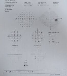
She came to the exam uncorrected with a VA of OD 20/70, OS 20/40, pinhole to OD 20/50 and no improvement in OS.

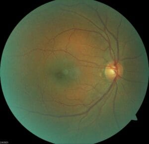
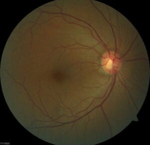
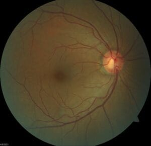
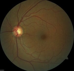
Anterior segment is stable to previous visits with clear PC-IOLs. Posterior segment shows stable moderate NPDR both eyes.

This doesn’t sound like anything unusual for this patient, we’d been monitoring her every six months for moderate NPDR since she first came to us in 2021. So why am I writing about her now?

For each of her prior visits, she had been able to see 20/25 or 20/30 with correction or pinhole. This was a change and decrease in vision, even though the patient herself had not noticed.

I get it. In a busy and crazy clinic like the community clinic I am in, it’s easy to just follow the plan and call it a day. But when something is different, it warrants digging a little deeper. So I had the student build the trial frame for this patient based on the prescription we found in January and her vision still did not improve.

The student was concerned that some of the hard exudates we saw were approaching the foveas and could be causing some macular edema so we took an updated OCT. At first glance, the student said everything looked fine. All retinal layers were intact and there was actually no evidence of macular edema. So if there wasn’t any edema and her refraction did not improve anything, why did this patient have reduced visual acuity?

Thankfully, when we looked at the OCT a little closer, we noticed actually a fair amount of vitreomacular traction in both eyes, enough to cause a little bit of distortion to the retinal layers, although she was correct, everything was still intact.

Now I felt a little better having a reason for this patient’s change in vision, but it got the student and I to have a conversation about how putting all this information together was important in helping us determine how to manage the patient moving forward. Did we need to refer her out to retina or could we still see her? Did we need to modify our follow up plan?

We opted to follow her a little more closely than 6 months and we’ll see how it goes from there.

Are there any takeaways or other points of discussion from you at this point? I’m always interested.

Patient case for August 18, 2023

Recently, we worked with a 55 year old black female who came in for her annual eye exam with no new complaints except she wanted to check on her eyes and get refills for her eye drops. She has been diagnosed with mild POAG both eyes and was scheduled to return for a visual field last year but no showed, so this is her first visit in about 1.5 years.

Systemically, she has a chronic hepatitis C infection with cirrhosis, arthritis, chronic back pain, and insomnia. She is taking latanoprost, timolol, cyclobenzaprine, diclofenac, melatonin, and trazodone.

The patient is correctable to 20/20 in each eye. Pupils, EOMs, and FDT visual field screener were clear today. She has some mild nuclear sclerosis and otherwise clear anterior segment. Posterior segment is also clear except for nerves with a C/D of 0.75 round in both eyes.

Historically, her max IOP was 16/18. In office today, IOP was 15/15.

Pachymetry is 517/518, gonioscopy showed open angles 360 OU.

An HVF 24-2 was last performed in 2021, which showed possible superior and inferior nasal steps both eyes after clear visual fields in 2019.

OCT of the RNFL both eyes showed thinning in the inferior-nasal quadrant for the right eye, thinning in the superior-temporal quadrant of the left eye.

Looking back at her visit history, she has had quite a few visits where she had cancelled them or no showed. At the end of the exam, she mentions she is always traveling and that’s why she can’t always come in. She also mentions that she skips a couple days’ worth of using her eyedrops when she’s waiting to pick up her new bottles from the pharmacy.

Given her appointment history and her IOPs, what would be your course of managing this patient next? I know I want to repeat her visual field for sure and check her IOP again. Is it worth it to consider adding another drop or change to a combo drop at the next visit? Is it worth it to refer her over to glaucoma to consider other interventions if her IOP still doesn’t go down?

What would you want to do for this patient?

Patient case for August 11, 2023

This week, we had a 54yo Filipino transgender female coming in for their first eye exam with us. They report distance blurred vision but no near problems, and they don’t have glasses.

Systemically, the patient has been diagnosed with DM2 and is s/p amputation of a toe on their left foot in July. They also have HTN, hypercholesterolemia, osteomyelitis, and history of a left sided stroke February of this year resulting in partial paresis on the right side and some residual speech difficulties. They have no medicinal allergies, and they report good compliance with medications: hydralazine, losartan, carvedilol, metformin, atorvastatin, aspirin, clopidogrel, glipizide, and insulin. Their last blood sugar was 120, but the latest HbA1c was 11.7 from February this year. They have an upcoming appointment with their PCP towards the end of this month and have been going through physical, occupational, and speech therapy since April.

Uncorrected visual acuity was OD 20/200, OS 20/125+1, with pinhole to 20/60, 20/50, respectively. Near VA was OD 0.4/1M, OS 0.4/0.63M.

Pupils, EOMs, and confrontation visual fields were full in both eyes.

The patient refracted to 20/40 OD with -2.75DS, OS 20/30 with -2.25 DS. There was no further improvement with pinhole.

Goldmann tonometry was 12/14 mm Hg. Anterior segment shows 1+ NS OU. Posterior segment was clear of any diabetic retinopathy or macular edema. There were no other remarkable ocular health findings.

What would you do in this case? The patient is certainly recovering from recent surgery still and still undergoing regular therapy for the sequelae of their stroke. Is there anything that could be contributing to this patient’s decreased visual acuity?

Would you prescribe glasses for this patient? How would you like to follow up with them?

Any other thoughts?

Patient case for August 4, 2023

A 60yo black male came in for a two month follow up after his comprehensive exam to repeat his visual field and check his IOP. Systemically, he is being treated for type 2 DM, sleep apnea, and POAG. His current medications include aspirin, Farxiga, metformin, pravastatin, Latanoprost and Cosopt.

His incoming vision is 20/20 in each eye and pupils and EOMs are normal in both eyes.

He has some mild nuclear cataracts as the only significant anterior segment finding. IOPs are 24/24 at 11:30 AM at this visit. Small pupil exam shows no evidence of drance hemorrhages and otherwise looks stable to previous photos as seen below.

His latest visual field is shown next.

There is no change compared to baseline field taken in 2018.

Other test findings include pachymetry of 514/526 and open gonioscopy both eyes. An RNFL OCT shows OD borderline thinning superior and OS thinning superior-temporally.

Prior to treatment, his max IOP was 25/25. The patient was treated with Latanoprost only for two years and during that time, IOPs ranged from 16-22 OD, 15-22 OS. Cosopt was added and IOPs have continued to range from 20-24 in both eyes in the last two years.

The patient admits to often missing his eyedrops a few times each week.

Given the lack of adequate IOP reduction but otherwise stable OCT and visual fields in this patient’s case, what would you do next? How would you classify this patient’s POAG? What are your comments on the visual fields?

Patient case for July 28, 2023

A 51 year old Hispanic female came in for her annual exam. She mentions she has some blurred vision after two hours of looking at her phone or doing close work. She closes her eyes to rest them and that makes things better. She doesn’t currently use glasses, though she has had them in the past.

Health wise, she is healthy without any medical conditions, has no allergies, and is not taking any medications.

Initial confrontation testing is all normal. She is correctable to 20/20- in each eye with a mild prescription of hyperopia and astigmatism. An appropriate add allows her to see 0.4/0.4M.

Anterior segment shows some mild interpalpebral staining and mild nuclear sclerosis in both eyes. IOPs are 18/18.

Peripheral posterior segment is unremarkable but posterior pole segment findings are seen as below.

Is anything remarkable here looking at the nerve, the blood vessels, the macula area?

The following are photos from an earlier visit in 2019.

Are there any differences?

I hope you can appreciate the significant loss of rim tissue in the left eye with the associated nerve fiber layer defect. Historically, this patient had been monitored for her large C/D’s over the years. Pachymetry is 456/450 nm, gonioscopy is open in both eyes, and a couple of 24-2 visual fields were clear. IOPs have ranged from 13-19 OD, 15-19 OS. She also had a drance heme of the inferior rim in the left eye in 2022. OCT progression analysis shows no statistical change in the right eye over the last 4 years, but significant thinning of the left inferior rim in that same period.

How would you manage this patient? If you are to medically treat, what’s your eye drop of choice given the monocular presentation of this condition? What’s your plan for the next follow up and moving forward?

Skip to content