Melissa Valdellon

Patient case for June 30, 2023

Today, let’s discuss this pediatric patient who came into the office for their first eye exam in the states after immigrating from Turkey. A previous eye doctor had let him know he had an eye turn and was given glasses. Mom says that the glasses helped the eye turn, but then the glasses were lost sometime in the last month. The patient is otherwise healthy, has no allergies, and is not taking any medications.

Pupils, confrontation fields, EOMs, and color vision are all normal. He did not appreciate randot stereoacuity and he reported 5 lights on worth 4 light.

Distance cover test reveals about 27pd left esotropia, whlie at near, it’s measured as 16pd left esotropia; the patient was uncorrected for both emasurements.

Uncorrected visual acuity is OD 20/20, OS 20/20.

Dry retinoscopy and refraction show:
OD: +0.75 DS  20/20-
OS: +1.25 -0.50 x 175  20/20

At this point, what drops would you use to dilate this patient and why? Can you review the different properties and effects of Tropicamide vs Cyclopentolate vs Phenylephrine?

~ ~ ~

We ended up using 1% Cyclopentolate for this patient and got the following results:
OD: +3.00 -0.50 x 180  20/20
OS: +3.75 – 0.50 x 170  20/20

All ocular health findings were normal.

What glasses prescription would you recommend for this patient? Given that the patient is 8 years old and able to see 20/20, what is your recommended follow up for this patient? If the patient were actually 5 years old, would any part of your management be different? And lastly, what are your thoughts around strabismus surgery in this case?

I’m looking forward to reading your thoughts.

Chocolate chia pudding

If you ask one of my students from the last couple years, they would probably let you know that most of the time, I bring lunch from home. And most of those times, it’s chia pudding.

Prepping this meal has become so second nature, I don’t have to think about it more than what extra I want to add each day, which also depends on what I have in my pantry on hand.

My basic recipe for this is:

  • 3 Tb chia seeds
  • 1 Tb sprouted quinoa
  • 1/3 to 1/2 Tb cacao powder
  • generous dash of cinnamon powder
  • 2 Tb mixed berries (usually goji and then maybe dried cranberries or raisins)
  • 2 Tb nuts or seeds (like sliced almonds or walnuts or pumpkin seeds)
  • 2/3 cup almond milk

I put this all the dry ingredients in a hydroflask first, add the milk last, and then and stir for a couple minutes, trying to break up all the chia chunks that can sometimes form. Then it’s done. It just sits in my bag until lunch time and it’s all ready. Just a couple more stirs to make sure the cacao powder is thoroughly mixed into the pudding and then it’s time to dig in.

I’ve swapped almond milk for coconut milk, taken out the cacao powder for dried coconut. I’ve added fresh berries too for some added sweetness when I don’t want the chocolate taste. All so good!

What’s your favorite chia pudding recipe? I’m always looking for new ways to have this for my work lunch days.

BIO tips for the straightforward patient

I’ve given a lot of mindset tips over the last few weeks regarding approaching clinic and your optometric education. Today, let’s talk about actual exam skills.

The last few years, it’s not an uncommon occurrence when a patient or a student makes a comment regarding my BIO “dance”. I always laugh and smile and say it’s all because of my years of doing yoga. It’s really hard for me to explain it, but my BIO flow involves a combination of slow movements, scanning in combination with lens tilting, and directing patient gazes that makes it work.

And it all evolved because of a couple of things. First, I remember that as a student, I continually got myself confused when I switched eyes in between scans. For example, doing all superior gazes of both eyes first, then all horizontal views, then all inferior views. My mind just couldn’t keep track of where lesions were. So rather than try to fight that, I learned that for me, scanning one eye at a time and recording those lesions first before going to the other minimized the chance of my recording a lesion in the incorrect eye.

Second, I quickly got very tired of the inefficiency with standing up and sitting down at a stool, adjusting its height every time I needed to get a new quadrant view. So my entire “routine” now is done standing with the patient seated and the seat usually all the way to the floor rather than raised to any height at all. Sometimes that means I’m tiptoeing a bit if a patient is particularly tall, or having that patient tilt their head ever so slightly inferior so I can sweep further out. For smaller kids, sometimes that means having the child stand on the edge of the chair and sometimes it means raising the chair so I’m more comfortable doing my sweep rather than crouching down as much.

Third, when I scan, my goal is to make a continuous sweep from the edge of the posterior pole at the level of the arcades all the way out to the periphery and sweep back in. To make sure I don’t minimize the chance of missing any peripheral findings, rather than doing a straight line sweep from arcades to periphery, I’ll make more of a flower petal shape coming back, moving my head a little bit so that the return sweep is covers new retina or overlaps slightly what had already been covered.

Fourth, I keep my light beam on its medium size setting and I use a good amount of light. Don’t think you’re being nice by using a dim light setting on your patients just to make sure they’re uncomfortable. Your job is to make sure their eyes are healthy and you don’t want to miss any subtle findings. On the flip side, don’t make the light so bright that they’re noticeably trying to squeeze their lids tight every time you approach their pupils with that light. I don’t like to fiddle with the light beam settings once the BIO is on my head and over the years, I’ve just found that a lot of glare is minimized simply by using the medium sized beam. And even if I have to use a brighter light, since I’m sweeping relatively quickly for my peripheral views and not lingering in any one spot too long, most patients are able to tolerate the time spent performing BIO.

Fifth, pay attention to where your beam and the patient gaze are and try to keep this as straight a line as possible. This ensures I’m getting the exact views I want. Simple in theory, but it took awhile for that to really lock in for me. It took an attending pointing out that while I had asked a patient to look superiorly, I was off to one side of a patient so my view was not the straight superior gaze I thought I was getting.

Lastly, keep practicing and keep adjusting. I know I spent many hours in pre-clinic practicing with classmates before we were ever in clinic and I still knew I had a ways to go. Have your attendings watch your BIO flow and give you feedback too. This is NOT an easy skill to master and it took a couple years of patient care to honestly feel like I wasn’t going to miss any major findings. And if your attending does point something out to you that you missed on your exam? Do NOT take it personally. Put your BIO back on after they leave and look for what you missed. After the patient has left, figure out how you can get to that spot in your next patient and patient after that consistently. You’re calibrating your sensitivity to spotting what’s normal and abnormal as well as learning how you need to move or place your hands and light in just the right spot so everything comes together for that perfect view. And just because what I shared with you today works for me doesn’t mean it has to work for you. Play with it. Have fun while you’re practicing.

We’ll talk about troubleshooting for the not so straightforward patient another time. Happy practicing!

Patient case for June 23, 2023

I am very grateful to work in a community clinic where I can speak with fellow doctors of different specialties about our mutual patients and they feel welcome to walk over anytime they want to discuss one of their patients with any eye concerns. This following case is just one recent example.

One of the clinic pediatricians came by asking if I could see one of her patients. The patient is a 13 year old female complaining of a sudden onset red right eye that she noticed when she first noticed upon waking up four days ago. Besides the redness, she says it felt like something was in her eye and irritating it. She reports that the eye has been watery with some discharge. She doesn’t remember anything getting into her eye, but she says she used some shimmery makeup two days prior and thinks maybe that could have irritated it.

After a day of no improvement, she went to the emergency room and was prescribed polytrim antibiotic eye drop. She reports good compliance with using the medication every 3 hours as prescribed but there has been no change in the redness or irritation and now the redness is starting to affect the left eye, though not as bad yet.

Overall, she says her eyes feel itchy and they burn. They don’t hurt really but she does have a small headache. And on further questioning, she reports that she hasn’t been recently sick and no one in her family and no one she’s been around in the last few days has been ill either.

Medically, the patient is very health with no conditions, no allergies, and is not taking any other medications besides polytrim eye drop.

Incoming visual acuity is 20/20-3 and 20/25+2 with easy pinhole to 20/20 each eye. Pupils and EOMs are normal both eyes.

Anterior segment exam findings shows clear lids and lashes, but conjunctival hyperemia, chemosis, mixed papillae and follicles OD worse than OS. There is patchy 1+ PEE peripherally on the cornea OD but otherwise clear centrally and no PEE in the left eye. Anterior chambers are deep and quiet, irides are brown and flat, lenses are clear, and vitreous is normal in both eyes. A small pupil posterior segment evaluation shows pink, distinct nerves with 0.40 cupping both eyes, shiny RNFL, normal vasculature, and flat and even macula of both eyes. IOPs are equal at 12/12 with Goldmann tonometry.

Given what you know, does this match the ER diagnosis of bacterial conjunctivitis? What in the exam findings suggest that this is bacterial in nature and what doesn’t?

What else is on your differential diagnosis list? How can you rule in or out allergic conjunctivitis? Viral? Iritis or uveitis? Do you think the patient’s headache is related to her ocular symptoms or not? What about her eye makeup use two days prior?

I can’t wait to read your thoughts!

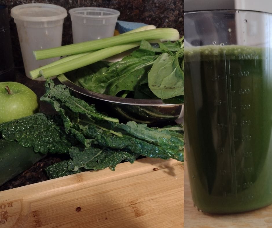
Apple, spinach, kale, cucumber, and celery juice

I really need to work on my photography skills – maybe that can be my next hobby to start!

Anyway, yay!!! For my birthday, I received a new cold press juicer. I loved my Greenstar, it got me through the beginning of my juicing journey, but this new Nama J2 has been amazing and I don’t have any excuse now to NOT juice. Now, as I said, I’m here to help you on your whole journey, including health. One of the things I can recommend is juicing. I was a skeptic too at first when I heard about it, but after doing a couple of juice cleanses over the past couple years, I can’t even begin to tell you how much lighter I feel, how much more clearer headed I am, and how grounded I feel when I do that.

Now, I’m not doing any cleanse at all, but I am really wanting to make juicing a part of my daily routine. I’m hoping this habit lasts because truly, every time I’ve been consistent with juicing in the past, I just feel great. And what’s not to love about increasing your daily intake of fruits and veggies?

Anyway, here’s a basic recipe that keeps me full and usually fills my 32oz water bottle for the day:

  • 2 green apples
  • half bunch spinach
  • 3 kale leaves
  • 2-3 ribs celery
  • 1 cucumber

Super easy, super basic, and not overwhelmingly “green-tasting” like other juices can be. Enjoy, and cheers to your health!

Letting go of expectations

I still remember the first patient I saw as a student clinician. The room was crowded because besides the patient and I, there was the attending doctor and 3 other classmates. We were doing team care for this young patient in his early 30s who came in complaining about irritated eyes.

I remember being so focused on ‘my part’ of the exam, I really didn’t pay as much attention to what was going on. I listened as the patient said he had no vision concerns, all he was worried about was his itchy, irritated eyes. I watched as a classmate did refraction and someone else did anterior segment. I did posterior segment and found nothing remarkable.

And we all got together with the attending doctor who confirmed our findings and did the final consultation. He agreed that the patient did not need glasses even though we had found a small prescription. All he recommended was over the counter allergy drops for his eyes and sent him on his way. Only then did I do a double take. Why? Because we weren’t giving the patient glasses.

Coming from someone who’s needed glasses since I was 6 years old, I obviously did not understand the concept that not everyone who went to the eye doctor would get glasses. Obviously, I knew that some people came in for other eye problems, but this was the first time it really hit me that even though this patient had a minor amount of refractive error, it wasn’t something to prescribe because he wouldn’t benefit from using it for his daily needs. I had to speak to the attending doctor about it afterwards and he was said that in his mind, why would he ask someone to pay good money for something they wouldn’t use? Yes, there were other kinds of doctors who would probably do the opposite and push and strongly recommend their patients to always obtain glasses to give themselves the best vision possible or latest fashion trend, regardless of actual need.

My attending that day, in a way, asked me what kind of doctor I desired to be. At that moment, I realized I had come into the profession with a preconceived idea of what an optometrist does. See a patient and give a prescription and maybe address some dry eyes. It was suddenly hitting me that there was more to my profession than just repeating a formula. What I was truly being asked was if I could provide personalized care to each and everyone of my patients. Sometimes, that would mean giving glasses. Sometimes that would not. Sometimes that would mean not seeing someone and referring them straight out to get other needs taken care of more urgently – it’s happened a few times already where a patient’s blood pressure is checked early in the exam and found to be emergency room level high and we stop an exam to get the person the more pressing care they need.

This exam showed me that some expectations should be let go. Rather than coming into a patient encounter expecting one thing, what is more beneficial is to be curious about what can come out of asking more questions, anticipating your findings, cross-checking to see if your guess matches results and determining why – why is the patient here, why are their eyes working and functioning and behaving the way they do, and why are they here in your chair at this moment?

When you let go of expectations and stay curious, you switch from technician mode who does everything according to a checklist to a patient advocate who partners with their patients in their overall care.

Outside the exam room and school setting, it wouldn’t be a bad idea to let go of expectations in other areas too. Can you identify where such thoughts are causing more stress than worth? Time to let it go… *cue Disney music here*

Patient case for June 19, 2023

Today’s patient is a 35 year old Hispanic male who is coming in for his annual exam. He is reporting some increased mild blur in his glasses, which he did not bring into the exam. He also wants to try contact lenses. He is otherwise healthy, has no allergies, and is not taking any medications.

Incoming visual acuity is OD 20/60, OS 20/50. Pupils, EOMs, and confrontation fields are all normal in both eyes.

Refraction results are:
OD: plano -4.00 x 017   20/20-
OS: -0.25 -2.75 x 159   20/20

Refraction results from two years ago showed 0.50D less cyl in the right eye but otherwise everything else was stable.

Anterior segment is clear and normal in both eyes, IOPs are 16/16, and posterior segment is normal with small nerves and small cups of 0.20 round OU, clear posterior poles, and stable lattice degeneration in each eye compared to 2 years prior.

How would you counsel this patient in terms of their wanting contact lenses? What would be your first choice to try? Is there additional testing you want to perform before trying lenses? Is there any hesitation recommendation contact lenses given he’ll be 40 in just a few more years?

Simple crudités and hummus

It’s finally kind of summer here in northern California. In the heat, that means it’s sometimes nice to have something a little lighter to munch on for dinner rather than prep and cook something hot.

One thing I’ve been trying to be more mindful of is purchasing produce that’s in season and locally grown. While I haven’t been able to make it a habit to visit my farmer’s market, I have been checking the tags more on my trips to Sprouts to see where my produce is from. For example, I love fresh tomatoes and I know they’re in season but after checking all the tags, who knew that the vast majority of tomatoes were shipped from Mexico?

Trying to be more conscious of my carbon impact has started to influence how I buy my groceries. I’m still working on general consumer products, but that’s another story.

Anyway, here’s a quick dinner I “came up with” when I was at the grocery store. I just got a tub of Sprouts organic hummus to go along with some fresh radishes and asparagus. I cleaned the asparagus and cut them into 2 inch long pieces. The radishes were sliced and the greens kept aside to add to my next day’s green smoothie.

I kept the veggies raw and just dipped them in the hummus for dinner. Overall, simple, delicious, and a different taste profile than I normally eat. Obviously, this can be made with any veggie and any type of hummus you prefer. Who knew crudités were more than just a party snack?

Happy nomming!

Keep a patient log

One of the simplest things I did as a student clinician was keep a patient log. Starting with the first patient I saw on my own, I kept track of certain facts about each patient – refractive error, best visual acuity, final prescription, ocular health findings, and – most importantly – any interesting pearls to take away from each patient.

Every patient, especially in the beginning of your clinical career, should have at least one thing you haven’t encountered before or have something that you didn’t understand. Those first few months, there should be a lot of “firsts”.

My first patient ever was a middle aged woman who needed to update her multi-focals. My attending and I spoke about the first instinct of just increasing an add to address her near blur as opposed to going over multi-focal designs, work environment and lighting and lifestyle needs to make sure her glasses were appropriately made. I have since had to adjust prescriptions for dental hygienists with decreased working distances, violin players who perform in in symphonies and need to see both the conductor in the distance and their sheet music up close, office workers who are on the computer long hours out of the day using multiple screens, and elderly patients who still sew and knit by hand.

I remember a male in his 40s, refracting one eye to an easy 20/20 but not the other eye. I did retinoscopy and refraction 3 times trying to get him to see better before moving on. After dilation, my attending took one look and ended up taking pictures – it turned out to be my first time seeing central serous retinopathy in person, explaining why this patient could not be refracted better than 20/60 in that other eye.

I remember a patient who had a moderately high myopic prescription who came in complaining that her vision was blurry with her new glasses. After checking their vision and refraction again, I found no change to the prescription. It wasn’t until another attending asked how the glasses were sitting on her face and demonstrated how adjusting the vertex distance that I realized how much of an impact that small detail could make because she was suddenly able to see much better and things seemed less distorted around her.

I wrote notes on how different doctors came up with a final prescription if an incoming visual acuity did not match refraction results. I wrote notes on how different doctors treated and managed their glaucoma patients. There were notes on how different types of urgent care patients were managed.

All of that went into a small notebook that I continued to add to until graduation, and then I hung onto it for a few more years afterwards even as a new doctor. It was always interesting to see what pearls I could take away.

Keeping a log is extra work, I know, but the ability to look back and see what you did for one patient can truly help you develop an understanding of how you can manage something similar when you see it again later. As an attending myself, I know I have spoken to more than one student about the benefits I got from it. One of them took it a step further and kept his log on google drive and then shared access with his attendings so we could give added input too for his patients. I thought this was brilliant – even if we couldn’t always go in depth about every single patient encounter, there were at least takeaway points from both his side and the doctors’ side for the student to review afterward.

Keep a log. In the end, it will also be a written testament of how much your clinical thinking has developed and how you’ve grown as a clinician in just two years.

Patient case for June 13, 2023

A 62 year old Hispanic female came in for her annual DM eye exam and was complaining of blurred vision after losing her glasses in Mexico a few months ago.

Medically, she is taking medications for mixed hyperlipidemia, osteoarthritis, type 2 DM, gastroesophageal reflux disease, and POAG with reported good compliance for everything. Her latest HbA1C was 7.4 and she did not check her blood sugar the day of her exam.

Incoming uncorrected visual acuity was 20/100 and 20/300 with pinhole to 20/30 and 20/50. Pupils and EOMs were normal.

Refraction results showed:
OD -22.5 DS  2025
OS -3.00 -0.50 x 170  20/40

Anterior segment showed meibomian gland dysfunction, nasal and temporal pterygium, 2-3+ NS, and vitreous syneresis, all in both eyes, though the cataract may have been slightly more hazy in the left than the right. IOP today was 14/14.

Posterior segment showed thin superior and inferior rims of both optic nerves with C/Ds of 0.65. There had been a hemorrhage on the inferior rim of the left optic nerve back in 2021. That has since resolved but there is now a corresponding mild nerve fiber layer defect. Otherwise, posterior segment was clear and without evidence of diabetic retinopathy.

For more background, this patient has been monitored for her nerve appearance since 2018. Pachymetry is 538/550. Gonioscopy is open in both eyes with no angle recession.

The patient has done multiple visual fields and for the right eye, there is a possible inferior nasal step that “comes and goes”, showing up in some fields but not all. The left eye also shows an inconsistent superior nasal step defect.

OCTs of the nerves show superior thinning that had been stable since 2018, but progressive inferior temporal thinning in the left eye more than the right since 2018.

Treatment of Latanoprost QHS was initiated in 2021 when the optic nerve hemorrhage was first seen. Prior to treatment, IOPs ranged from 19-22 OU. After treatment, her IOPs have fluctuated from 14-22, achieving IOPs of 15 or lower in half the visits since treatment was initiated.

Sorry for the lack of imaging to go along with the case but hopefully you were able to follow along. What would you prescribe for this patient for their refractive error or how would you counsel them in regards to their vision? What would you tell the patient and their doctor about their diabetes management? And what would you like to do about this patient’s glaucoma?

I look forward to hearing your thoughts.

Skip to content